
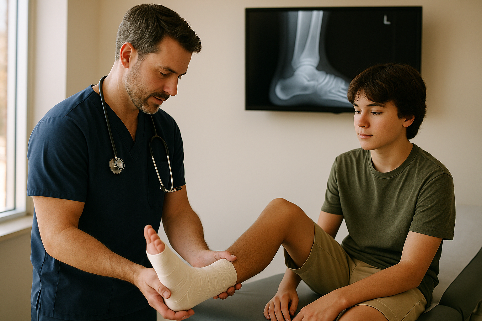
An ankle rolls on a curb, a soccer field, or the last step of a staircase. Within an hour it is swollen and purple and hurts to look at. The question every patient asks, usually before they have finished sitting down, is whether it is broken.
The honest answer is that swelling and bruising do not tell you. A bad sprain can look dramatic and a small fracture can look unimpressive. What does help is a set of clinical rules that have been studied for decades specifically to answer this question, and they perform remarkably well.
Do I need an X-ray for my ankle injury?
Under the Ottawa Ankle Rules, an X-ray is indicated if there is pain in the malleolar zone and any one of: bone tenderness along the distal 6 cm of the posterior edge of the tibia or the tip of the medial malleolus; bone tenderness along the distal 6 cm of the posterior edge of the fibula or the tip of the lateral malleolus; or inability to bear weight for four steps both immediately after the injury and at the time of assessment. In a pooled analysis the rules showed about 97 percent sensitivity for ruling out significant ankle fractures.
We care for patients from Bolingbrook and the surrounding communities including Romeoville, Woodridge, Naperville, Plainfield, Downers Grove, Lockport, Lisle, Darien and Lemont. Our clinic is at 148 S Bolingbrook Dr, open 7 days a week from 8 AM to 8 PM, with walk-ins welcome. If you are dealing with a sprain or possible fracture, you can walk in or book a visit online.
Why swelling is a bad guide
Soft tissue responds to injury with blood flow and fluid, and it does so enthusiastically. A completely torn ligament with no broken bone can produce spectacular swelling and bruising that tracks down into the foot over a couple of days. Meanwhile a small avulsion fracture, where a fragment of bone is pulled off by a ligament, can look almost boring.
This is why clinicians do not decide on imaging by looking. They decide by pressing on specific bones and by asking whether the patient can walk. Both of those turn out to carry far more information than appearance.
The Ottawa Ankle Rules, in plain language
The Ottawa Ankle Rules were developed to reduce unnecessary X-rays without missing fractures that matter. They are among the most validated decision tools in emergency medicine, and they are deliberately built to err toward imaging when there is doubt.
- 1Start with the malleolar zone
If there is no pain around the bony bumps on either side of the ankle, the rule does not apply and imaging is generally unnecessary. - 2Press along the back edge of the inner ankle bone
Bone tenderness along the distal 6 cm of the posterior edge of the tibia, or at the tip of the medial malleolus, is a positive finding. - 3Press along the back edge of the outer ankle bone
Bone tenderness along the distal 6 cm of the posterior edge of the fibula, or at the tip of the lateral malleolus, is a positive finding. - 4Ask about weight bearing
Inability to bear weight for four steps, both immediately after the injury and at assessment, is a positive finding. Limping counts as bearing weight. - 5Any one positive finding means X-ray
Pain in the malleolar zone plus any single one of the three findings above means imaging is indicated.
The word doing the heavy lifting is bone. The rule asks about tenderness directly over bone, not general soreness in the surrounding soft tissue. An ankle that is diffusely tender across the swollen outer side but not specifically tender on the bony landmarks behaves very differently under this rule than one that hurts sharply at the tip of the malleolus.
How well the rules actually perform
This is where the evidence is worth quoting rather than paraphrasing, because the two numbers point in different directions and both matter.
Sensitivity is the ability to correctly identify injuries that do have a fracture. Specificity is the ability to correctly identify those that do not. High sensitivity with low specificity is exactly what a safety screening tool should look like: it very rarely misses a fracture, at the cost of sending some people for an X-ray that turns out normal. That trade is intentional.
Data: Systematic review and meta-analysis, diagnostic accuracy of the Ottawa ankle rule in adults (PMC)
In practical terms, a negative result is highly reassuring. A positive result means an X-ray is warranted, not that a fracture is likely. Most people who meet the criteria and get imaged do not have a broken bone, and that is the rule working as designed.
The rules have also been studied in younger patients. Research supports the Ottawa Ankle Rules as a reliable clinical instrument for detecting fractures in children and adolescents, which matters in a clinic that sees a lot of school sports injuries. That said, growing bones have features adult bones do not, including growth plates that can be injured in ways that need a clinician’s judgment rather than a checklist.
What an X-ray shows, and what it misses
A frequent misunderstanding is that a normal X-ray means nothing is wrong. It does not. It means no fracture was seen, which is a narrower statement.
| Shows well | Shows poorly or not at all |
|---|---|
| Most fractures and bone fragments | Ligament tears, which are soft tissue |
| Bone alignment and joint dislocation | Tendon injuries |
| Some foreign bodies, such as glass or metal | Cartilage damage |
| Changes in bone from older injuries | Some hairline and growth-plate injuries in the first days |
This is why a normal X-ray is often followed by a diagnosis of a severe sprain, and why a clinician may recommend re-examination in a week if pain and tenderness are not following the expected path. Some injuries only become visible on imaging after a few days.
Caring for a sprain that does not need imaging
For sprains that do not meet imaging criteria, management is unglamorous and effective. The standard approach is a RICE plan, meaning rest, ice, compression and elevation, in the early days after injury.
- Rest relatively rather than absolutely. Protect the joint early, then begin gentle movement as pain allows rather than immobilizing for weeks.
- Ice for short intervals rather than long ones, with a barrier between the ice and the skin.
- Compression with an elastic wrap applied snugly but never tightly enough to cause numbness, tingling or colour change in the toes.
- Elevation above the level of the heart where practical, which does more for swelling than most people expect.
The single best predictor of a good outcome is not the first 48 hours, it is what happens in weeks two through six. Ankles that are never rehabilitated tend to become ankles that roll again, because the ligaments heal but the balance and strength do not come back on their own. If you take one thing from this section, make it that.
When to skip urgent care and go to the emergency room
Some injuries are past what a walk-in clinic should handle, and it is better to know before you drive.
- Obvious deformity, where the joint or limb is visibly out of position
- Bone visible through the skin, or an open wound over a suspected fracture
- A foot or toes that are numb, cold, pale or blue, which suggests a problem with blood supply or nerves
- Inability to move the foot or toes at all
- A high-energy injury such as a significant fall, a car crash or a crush injury
- Severe pain that is out of proportion to the appearance of the injury, particularly with a tight, tense swollen compartment
Everything else, the rolled ankles, the jammed fingers, the wrist that took a fall, the foot that got stepped on in a game, is well within the range of a clinic with imaging on site.
Why on-site X-ray changes the visit
The practical advantage of having X-ray in the building is that the question gets answered in one stop. A clinician examines the ankle, applies the criteria above, images it if indicated, and reads the result during the same visit. You leave knowing whether a bone is broken, with a wrap or a boot if needed, and with instructions.
Without on-site imaging, the same injury becomes an examination, a referral to an imaging center, a second trip, and a wait for someone to call you with the result. For a swollen ankle in August, that is the difference between an afternoon and a week.
We have X-ray on site at our Bolingbrook clinic, with splints and crutches, and see patients from across the surrounding communities seven days a week, 8 AM to 8 PM. We do not offer CT or MRI, so if an injury needs advanced imaging we will tell you and help you get there rather than pretending a plain film settles it.
The same logic applies to knees
Ankles are the most common version of this question, but knees generate it too, and there is a parallel decision tool with the same design philosophy.
| Criterion | What it means |
|---|---|
| Age 55 or older | Age alone is a criterion in this rule. |
| Isolated patellar tenderness | Tenderness on the kneecap with no other bony tenderness around the knee. |
| Fibular head tenderness | Tenderness at the bony prominence on the outer upper leg, roughly 1 to 2 cm below the joint line, not at the joint line itself. |
| Inability to flex to 90 degrees | Assessed by asking the patient to actively bend the knee during examination. |
| Inability to bear weight for 4 steps | Two steps per foot, both immediately after the injury and at assessment. Limping counts as bearing weight. |
As with the ankle rule, the criteria are deliberately broad so that fractures are not missed. Meeting a criterion means imaging is warranted, not that a fracture is likely.
The knee rule has been validated in adult populations, and its purpose is identical: reduce imaging that will not change management, without missing the fractures that will.
How sprains are graded, and what each grade means for you
When imaging is negative and the diagnosis is a sprain, the follow-up question is how bad. Clinicians generally describe ligament injuries in three grades, and the grade shapes the timeline more than anything else.
| Grade | What has happened | What it usually looks like |
|---|---|---|
| Grade 1 | Ligament fibres stretched, with microscopic tearing | Mild swelling and tenderness, joint feels stable, walking is uncomfortable but possible |
| Grade 2 | Partial tear of the ligament | More swelling and bruising, noticeable looseness on examination, walking is difficult and painful |
| Grade 3 | Complete tear of the ligament | Significant swelling and bruising, marked instability, often unable to bear weight; sometimes less painful than a grade 2, which is counterintuitive |
Grade 3 injuries can hurt less than grade 2 injuries because a completely torn ligament stops generating tension. Pain level is therefore a poor guide to severity, which is another reason examination matters more than how it feels.
Recovery time scales with grade. A grade 1 sprain often settles within a couple of weeks. A grade 2 typically takes several weeks. A grade 3 can take months and sometimes involves orthopedic referral, particularly when the joint remains unstable.
Other injuries we image on site
Ankles get the attention, but the same one-visit logic applies across most of what walks into an urgent care after a game or a fall.
- Wrists after a fall onto an outstretched hand, which is the classic mechanism for a distal radius fracture and also for a scaphoid injury that can be subtle on early films
- Fingers and thumbs jammed in ball sports, where the question is usually fracture versus dislocation versus ligament injury
- Feet and toes, including the very common stepped-on foot and the less common but more important midfoot injury
- Elbows and forearms in children, where growth-plate injuries need a careful eye
- Ribs after a fall or a direct blow, where imaging is often more about ruling out complications than about the rib itself
- Knees and lower legs following the criteria above
A note on wrists specifically. A scaphoid fracture, in the small bone at the base of the thumb, is notorious for not showing on an initial X-ray. If tenderness is in that specific spot, a clinician may treat it as a fracture and re-image later even when the first film looks normal. That is deliberate caution, not indecision, because a missed scaphoid fracture can have real long-term consequences.
Rehab is the part that prevents the next one
The single most useful thing in this article for anyone who has already sprained an ankle: the injury that predicts your next ankle sprain most strongly is your last ankle sprain.
Ligaments contain sensors that tell your brain where the joint is in space. Injury damages that feedback along with the tissue, and it does not fully return on its own just because the swelling went down and walking stopped hurting. Restoring balance and strength deliberately is what closes the loop.
- Early on, protect the joint and control swelling, but begin gentle movement as pain allows rather than immobilizing for weeks.
- As pain settles, work on range of motion in all directions.
- Add strengthening, particularly of the muscles running along the outside of the lower leg.
- Add balance work. Standing on the injured leg, eventually with eyes closed or on an unstable surface, directly retrains the sensors that were damaged.
- Return to sport progressively rather than in one step, and consider bracing or taping for the first season back in cutting sports.
If pain, swelling or instability is not steadily improving over a few weeks, that is worth another look. Persistent symptoms after what was diagnosed as a simple sprain sometimes reflect an injury that was not visible initially, and that is exactly the situation where re-examination earns its keep.
This article is for general education and is not medical advice. It does not replace an evaluation by a licensed clinician. If you think you are having a medical emergency, call 911 or go to the nearest emergency room.
Frequently asked questions
Can I walk on it if it is broken?
Sometimes, yes. Being able to walk does not rule out a fracture, which is why the Ottawa Ankle Rules combine weight bearing with specific bone tenderness rather than relying on walking alone. Limping counts as bearing weight.
How accurate are the Ottawa Ankle Rules?
In a pooled analysis in adults the rules showed sensitivity of about 97.3 percent and specificity of about 36.6 percent. That means they rarely miss a significant fracture, at the cost of some normal X-rays.
Does a normal X-ray mean nothing is wrong?
No. It means no fracture was seen. X-ray does not show ligament, tendon or cartilage injuries well, and some hairline and growth-plate injuries are not visible in the first few days. A severe sprain can be a genuinely significant injury.
Should a child’s ankle injury be handled differently?
The rules have been supported as reliable in children and adolescents, but growing bones have growth plates that can be injured in ways a checklist does not capture, so a clinician’s assessment matters more in younger patients.
Do you have X-ray on site?
Yes. We have X-ray and labs in the clinic, so a provider can examine the injury, image it if indicated and review the result during the same visit. We do not offer CT or MRI.
Sources
- Diagnostic accuracy of the Ottawa ankle rule in adults: systematic review and meta-analysis (PMC)
- Ottawa Ankle Rules in children and adolescents (PMC)
- MDCalc: Ottawa Ankle Rule
- American Academy of Pediatrics: HealthyChildren.org
Guidance from public health agencies changes. Dates and figures above reflect the sources as published at the time of writing. Always confirm current requirements with the issuing agency or your school district.
Need care today in Bolingbrook?
Walk in or book online. Open 7 days a week, 8 AM to 8 PM, with X-ray and rapid tests on site.